.svg) info@scarsandlasers.co.nz
info@scarsandlasers.co.nz
15 Green Lane East, Remuera, Auckland, New Zealand

Monday - Friday: 7:45 am - 5:00 pm

Squamous cell carcinoma (SCC) is the second most common form of skin cancer after basal cell carcinoma (BCC). It develops from squamous cells, which make up the surface layer of the skin (the epidermis).
Squamous Cell Carcinoma is the second most common form of skin cancer after Basal Cell Carcinoma. It develops from squamous cells, which make up the outer layer of the skin (epidermis). SCC typically arises in areas that have experienced long-term sun exposure, but it can also occur on parts of the body not typically exposed to sunlight.
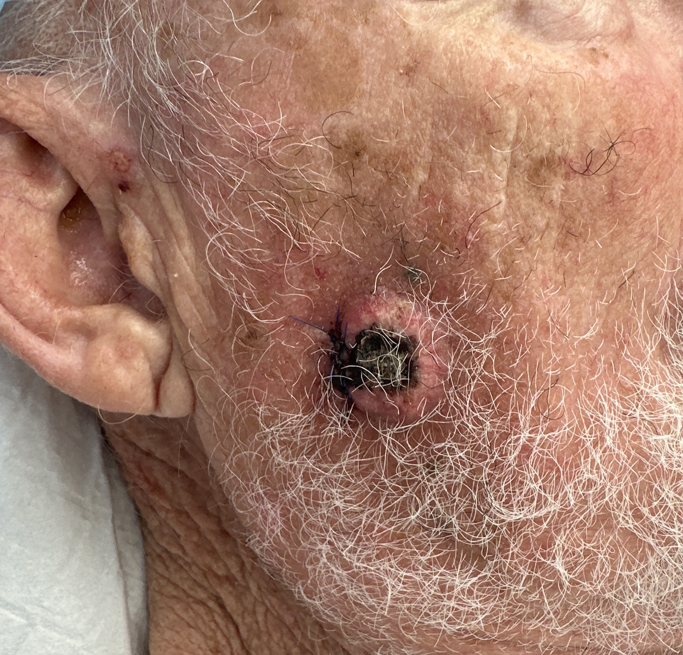

Squamous cell carcinoma (SCC) is a common skin cancer that begins in squamous cells, which are found in the outer layers of the skin. It often develops on sun-exposed areas such as the face, ears, scalp, neck, forearms, and backs of the hands, but it can occur anywhere on the body. SCC can also occur in special sites such as the lip, inside the mouth, around the genitals/anus, or near/under a nail. Many SCCs are treated successfully, especially when found early. However, SCC is not something to watch and wait on. Some SCCs can grow deeper into the skin, damage nearby tissues, and a smaller proportion can spread (metastasise) to lymph nodes or other areas. If you have been told you have SCC, your clinician will usually describe it as 'low-risk' or 'high-risk' based on the biopsy features, the size, and the location. That risk level helps guide how wide margins should be, whether Mohs surgery is appropriate, and how closely follow-up should be arranged.
SCC can look different from person to person. It commonly appears as a persistently scaly, crusty, or thickened area of skin, or as a firm bump that may feel rough. It may ulcerate (break down), ooze, or bleed, and it can be painful or tender although some lesions are painless. Common warning signs include: A sore that does not heal, or heals and then comes back.
A rough, scaly patch that persists and gradually enlarges.
A raised growth with a central dip, crust, or ulcer.
A spot that bleeds with minor knocks or when washing your face.
A lesion that is becoming more painful, numb, or tingly.
A change on the lip (persistent scale, thickening, or ulcer) or a new lesion under/around a nail.
Because SCC can mimic other skin conditions, the most reliable way to know what it is is a clinical assessment and, when appropriate, a biopsy. If something is new, changing, or not healing over a few weeks, it is worth getting checked.
Most SCCs are cured with appropriate treatment. The seriousness depends on 'risk features' that increase the chance of the SCC returning after treatment or spreading to lymph nodes. Factors that can increase risk include (your clinician will interpret these for your case): Location (some areas on the head and neck are considered higher risk, such as the ear and lip).
Size (larger tumours generally carry higher risk) .
Depth of invasion and certain biopsy features such as poor differentiation or perineural involvement.
Recurrence (a tumour that has been treated before and has returned).
Reduced immune function (for example, organ transplant recipients or people on long-term immunosuppressive medicines).
If there are concerning features, your clinician may discuss additional steps such as examining nearby lymph nodes, arranging imaging, or coordinating care with other specialists. Not everyone needs these steps - it is tailored to the individual risk profile.
SCC is most commonly driven by long-term ultraviolet (UV) exposure from the sun and/or tanning beds, which can damage DNA in skin cells over time. It can also arise in areas of chronic inflammation or injury (such as longstanding ulcers or scars), and other factors may contribute depending on the site.
Risk tends to be higher if you:
Have had significant sun exposure over many years or frequent sunburns.
Use (or used) tanning beds.
Have had previous skin cancers or many sun-damaged 'pre-cancer' spots (actinic keratoses).
Have a weakened immune system (for example after an organ transplant or due to certain medications).
Have SCC on the lip, ear, or certain other higher-risk sites SCC can occur in people of all skin tones.
In darker skin tones, SCC is more likely to occur in areas of chronic inflammation or scars and may not be sun-related. That is one reason clinicians take non-healing or changing lesions seriously regardless of skin type.
SCC is diagnosed with a skin biopsy. A biopsy is a quick procedure performed under local anaesthetic, where a small sample (or sometimes the whole lesion) is removed and examined under a microscope by a pathologist. The biopsy confirms whether it is SCC and provides detail about the type and risk features. Common biopsy types include a shave biopsy, punch biopsy, or excisional biopsy. Which one is used depends on the location, size, and the clinicians judgement. After the biopsy you will have a small wound that needs simple wound care for several days. Your pathology report may include information such as tumour thickness/depth, degree of differentiation, and whether there is perineural involvement. These details help guide treatment choice (standard excision vs Mohs vs other treatments) and follow-up planning.
Treatment is chosen based on the SCCs location, size, biopsy features (risk level), whether it is primary or recurrent, and your overall health. For many SCCs, surgical removal is the main treatment because it physically removes the cancer and allows margin assessment. Common treatment options your clinician may discuss include:
Standard surgical excision (cutting out the SCC with a margin of normal-looking skin)
Mohs micrographic surgery (layer-by-layer removal with immediate microscopic margin checking)
Curettage and electrodesiccation (scraping and cauterising) for selected low-risk lesions in suitable locations
Radiation therapy when surgery is not suitable or as an additional treatment in specific situations.
For advanced cases, referral for specialist systemic treatments (for example, immunotherapy) may be discussed
Your clinician will aim for a plan that provides a high cure rate while considering functional and cosmetic outcomes, especially for the face and other sensitive areas. They will also explain expected recovery time, scarring, and follow-up.
Mohs micrographic surgery (often called 'Mohs surgery') is a specialised technique that removes skin cancer in thin layers. After each layer is removed, it is processed and examined under a microscope on-site. The surgeon maps where any cancer cells remain, and only removes further tissue precisely where cancer is still present. This continues until all margins are clear.Compared with standard excision, Mohs offers two major advantages in the right setting:
Margin confidence: Mohs examines the tissue margins during the procedure, so the surgeon can confirm clearance before reconstructing the wound.
Tissue-sparing: Because only the areas with remaining cancer are removed, Mohs can preserve more healthy skin especially valuable on the nose, eyelids, lips, ears, and hands. Technically, Mohs uses a tissue processing method that allows a very high proportion of the peripheral and deep margins to be examined. This approach is one reason Mohs is often chosen for cancers in cosmetically or functionally important sites.
Mohs is often recommended when an SCC has features that make it more likely to recur or when precise margin control is important. It is commonly used for SCCs in high-risk areas (especially parts of the head and neck), for recurrent tumours, and for tumours with less well-defined borders. Mohs may be considered if your SCC is:
Many low-risk SCCs are safely treated with standard excision. Your clinician will recommend Mohs when the benefits (precision, tissue preservation, reduced recurrence risk) outweigh the additional time and complexity.
Mohs is performed as an outpatient procedure under local anaesthetic. Oral sedation is offered if requested. The area is numbed so you should not feel sharp pain during the procedure, but you will feel pressure or movement. If you are uncomfortable at any point, tell the team - more anaesthetic can usually be given.
A typical Mohs day includes:
Because the number of stages varies, the total time can range from a few hours to most of the day. It is best to keep the day flexible, bring something to do while waiting, and arrange transport if you think you may feel tired afterwards.
After SCC surgery, some swelling, bruising, and tenderness are common for the first few days. The exact recovery depends on the size and location of the wound and how it was repaired. Your clinic will provide wound-care instructions (cleaning, dressings, ointment, activity limits, and when stitches need review or removal).
How the wound is closed can include:
All procedures can leave a scar, but scars typically improve over months. Scar appearance depends on individual healing, wound tension, and location. Sun protection during healing is important because UV exposure can darken scars and slow improvement.
Possible risks to discuss with your clinician include:
Follow-up is tailored to risk. Some guidelines note that after complete excision of a low-risk SCC, no additional specialist follow-up may be required beyond routine skin checks, whereas higher-risk SCCs may need closer surveillance. Regardless of risk level, many people who have had one SCC are more likely to develop another skin cancer in the future, so ongoing self-checks and regular professional skin checks are important.
Practical prevention steps include: daily broad-spectrum sunscreen, protective clothing and hats, avoiding midday sun where possible, and avoiding tanning beds. Your clinician can also advise on managing sun-damaged skin and pre-cancerous lesions.
ACC funding eligibility is determined by the Accident Compensation Corporation on a case-by-case basis. Approval is dependent on injury acceptance, clinical indication, and ACC policy at the time of application. Not all treatments offered at Scars & Lasers are ACC-funded. Private fees may apply for unfunded components of care.